Why Cryo-EM Is Becoming One of Science’s Most Useful Vision Tools
For centuries, humanity's understanding of the world has been profoundly shaped by our ability to see it, and science is no exception. From Galileo's telescope revealing distant celestial bodies to the optical microscope unveiling the hidden world of microorganisms, new vision tools have consistently sparked revolutions in thought and discovery. Today, a powerful new lens is rapidly reshaping our view of the molecular universe: cryo-electron microscopy, or cryo-EM.
Cryo-EM is not just another imaging technique; it’s a paradigm shift in structural biology and beyond. It offers scientists an unprecedented look at the intricate machinery of life and matter, providing near-atomic resolution views of proteins, viruses, and complex molecular assemblies that were previously almost impossible to visualize in their native states. This capability is turning high-resolution visualization into a practical decision-making tool, fundamentally changing how researchers approach problems in drug discovery, disease understanding, and materials innovation.
How Cryo-EM Works Its Magic
At its heart, cryo-EM tackles a fundamental challenge: how do you image delicate biological samples at extremely high resolution without damaging them? Traditional electron microscopy often requires samples to be dried or stained, which can distort their natural structure. X-ray crystallography, another powerful technique, requires samples to be coaxed into forming perfect crystals – a challenging and often impossible task for many important molecules, especially large complexes and membrane proteins.
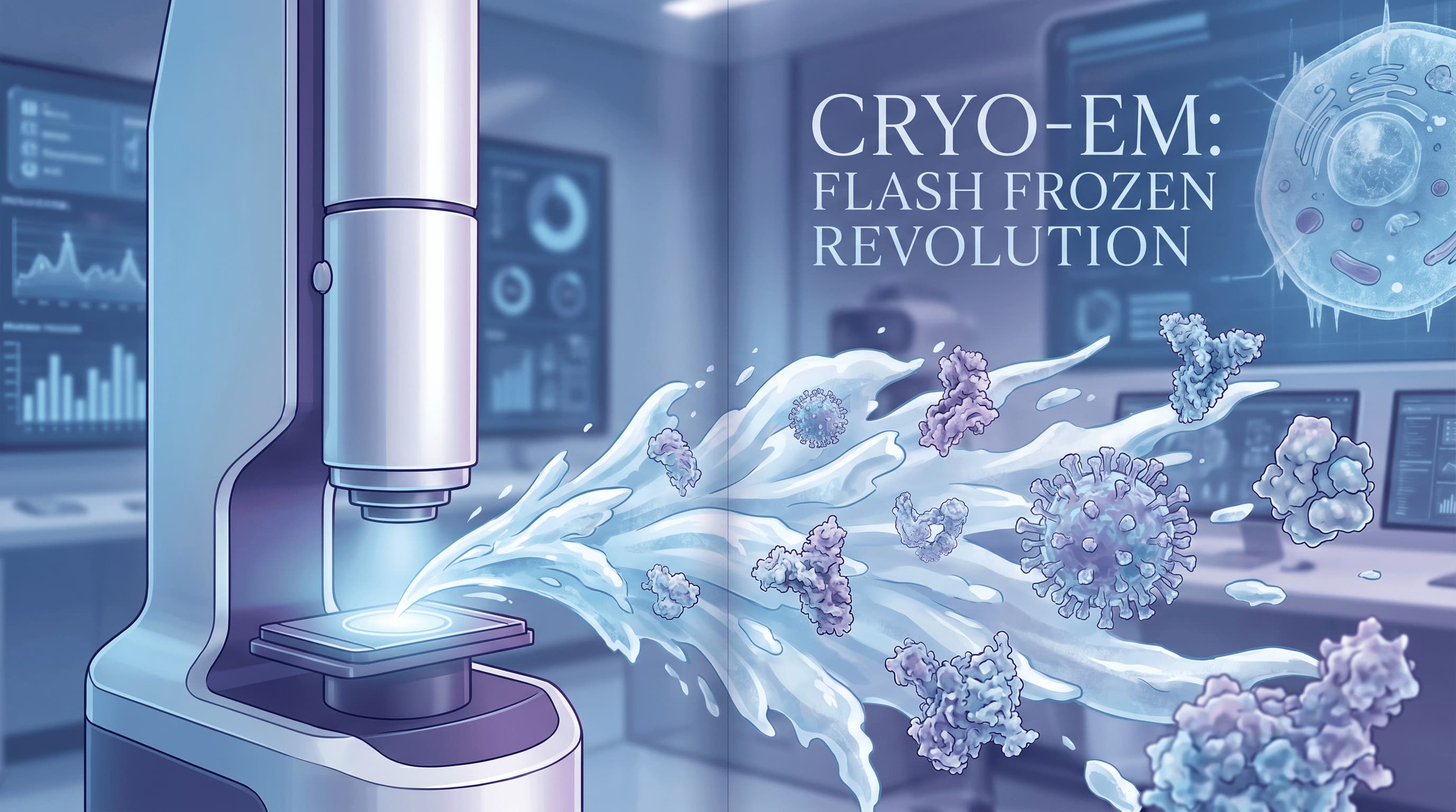
Cryo-EM circumvents these issues with an elegant solution: flash-freezing. Researchers rapidly plunge a thin layer of the sample solution into a super-cooled liquid, typically ethane. This process is so fast that water molecules don't have time to form destructive ice crystals; instead, they solidify into a non-crystalline, glass-like state called vitreous ice. This preserves the molecules in their natural, hydrated environment, essentially trapping them in a snapshot of their functional conformation.
Once frozen, these samples are then placed into a powerful electron microscope. Instead of a single image, the microscope captures thousands upon thousands of two-dimensional projections of the molecules from various orientations. Electrons, with their much shorter wavelength than light, allow for far greater resolution. Sophisticated computational algorithms then take these numerous 2D images and stitch them together, much like creating a 3D model from countless photographs taken from different angles. The result is a highly detailed, three-dimensional reconstruction of the molecule, often revealing features down to individual atoms.
Overcoming the Bottleneck: Why Cryo-EM is a Game Changer
Beyond Crystallization: Seeing the Unseeable
One of cryo-EM’s most significant contributions is its ability to bypass the "crystallization bottleneck" that has long plagued X-ray crystallography. For many crucial biological targets – particularly large, flexible protein complexes, membrane proteins embedded in lipid bilayers, and transient states of molecules – forming well-ordered crystals is exceedingly difficult or impossible. These are precisely the molecules often involved in critical cellular processes and disease pathways.
By eliminating the need for crystallization, cryo-EM has opened up vast new frontiers. Scientists can now visualize structures that were previously intractable, providing insights into mechanisms of action for drug targets, understanding viral replication, or deciphering the intricate machinery of cellular communication. This means we can now study molecules in states closer to their natural biological environment, yielding more physiologically relevant information.
Broader Applications: From Biology to Materials Science
While cryo-EM has revolutionized structural biology, its utility extends far beyond. Researchers in chemistry and materials science are increasingly leveraging the technique to understand complex structures, dynamics, and interactions in challenging samples. Imagine studying the precise arrangement of atoms in a novel catalyst, the defects in a semiconductor material, or the assembly of synthetic nanoparticles. Cryo-EM offers a unique window into these systems, providing structural information that can guide the design and optimization of new materials and chemical processes.
Guiding Discovery: A Practical Decision-Making Tool
The core power of cryo-EM lies in its ability to transform high-resolution structural data into actionable knowledge. When scientists can see the precise shape of a protein binding site, they can rationally design drugs to fit it. When they understand how a virus assembles, they can develop strategies to disrupt it. This direct visual evidence empowers researchers to make informed decisions, accelerating the iterative process of hypothesis, experiment, and refinement. It’s about more than just seeing; it’s about seeing clearly enough to build, modify, and innovate.
The Road Ahead: Challenges and Considerations
Despite its revolutionary capabilities, cryo-EM is not a universal panacea, and its implementation comes with significant considerations. It's important to approach this powerful tool with a balanced perspective, acknowledging its limitations alongside its strengths.
High Costs and Specialized Expertise
The initial investment for a state-of-the-art cryo-EM facility is substantial, often running into several million dollars for the electron microscopes themselves, specialized sample preparation equipment, and high-performance computing infrastructure. Beyond the hardware, operating a cryo-EM lab requires highly specialized expertise. Sample preparation, microscope operation, and particularly the complex computational data processing and 3D reconstruction demand extensive training and experience. This means that while the technology is transformative, access remains concentrated in well-funded institutions and specialized core facilities.
Throughput and Sample Preparation Nuances
While cryo-EM has made incredible strides in efficiency, it can still be a lower-throughput method compared to some other screening techniques, especially when dealing with a vast library of compounds or samples. Each sample requires meticulous preparation, and even with automation, the process can be time-consuming. Achieving the ideal vitreous ice layer with a perfectly dispersed sample is an art form, and sample quality remains paramount for obtaining high-resolution data. Impurities, aggregation, or insufficient concentration can all compromise results, making sample optimization a critical and often challenging step.
Data Analysis Complexity
The sheer volume and complexity of the data generated by cryo-EM experiments are immense. Processing thousands of 2D images, aligning them, classifying different molecular conformations, and reconstructing a high-resolution 3D map requires significant computational power and sophisticated software algorithms. Interpreting these maps, validating their accuracy, and extracting meaningful biological or material insights also demands a deep understanding of structural biology and computational methods. This data analysis bottleneck can be as challenging as the experimental work itself.
Conclusion: A Sharper Vision for Science
Cryo-EM has undeniably earned its place as one of science's most useful vision tools. By offering unprecedented glimpses into the molecular world, it has accelerated our understanding of fundamental biological processes, paved new avenues for drug discovery, and opened doors for innovation in materials science. While challenges related to cost, expertise, and data analysis persist, the ongoing advancements in instrumentation and computational methods are continually expanding its accessibility and capabilities.
As researchers continue to harness the power of cryo-EM, we can anticipate even more profound discoveries. It’s a testament to human ingenuity that we can now peer into the atomic architecture of life itself, transforming what was once invisible into a clear, actionable vision for the future of science and technology.